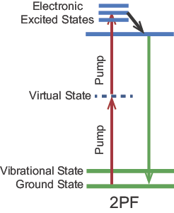
Two-Photon Fluorescence Microscopy

Conventional wide-field microscopy provides micron-level resolution laterally across a sample. In contrast, resolution into the depth of the sample, sometimes notated as the Z direction, can be considerably poorer due to signals that originate from various depths within a sample leading to obscuration of its origin. In a confocal laser scanning microscope, the fluorescence generated from a focused laser is imaged onto a pinhole, or confocal aperture, which blocks the fluorescence that is generated above or below the focal plane before reaching the detector. The intensity of the fluorescence is then measured as the beam is scanned horizontally across the sample to build up a 2D image. Finally, the sample is sequentially moved in the Z direction, allowing for 3D image construction with a spatial resolution close to the diffraction limit. This technique of confocal laser scanning microscopy dates back to 1969.

Two-Color (Non-Degenerate) Two-Photon Fluorescence Imaging

Confocal microscopes are essential tools in today's biomedical research laboratories, but they do have limitations. Due to losses at the pinhole and scattering of the excitation light, confocal microscopy techniques are best applied to thin specimens (< 20 µm). While the out-of-focus excitation does not contribute to the 3D image, it does increase the likelihood of photodamage in living specimens. In addition to the potential photodamage caused by typically-used VIS and UV wavelengths, they also do not penetrate deeply into tissue due to scattering. In 1990, workers at Cornell University demonstrated the use of two-photon excitation with laser scanning microscopy, denoted here as 2PF microscopy. In 2PF, two photons are simultaneously absorbed to cause a higher energy electronic transition in a fluorescent molecule as shown in Figure 1.
Energy level diagram showing 2PF of a fluorescent dye
Figure 1. Energy level diagram showing 2PF of a fluorescent dye.
>Since the nonlinear process of 2PF has a low cross-section for excitation, a high peak power laser source is required as well as a high NA microscope objective. This combination produces a small focal volume where the intensity is high enough to drive the 2PF process. The NIR wavelengths required for 2PF are about twice as long as those used in conventional confocal microscopy and have the important benefit of reduced scattering and thus deeper penetration. To achieve the high peak power required for 2PF without the high average power that would damage the sample, mode-locked fs lasers are required (see Methods for Pulsed-Laser Operation for details). A typical laser for 2PF microscopy produces 120 fs pulses every 12 ns (or a repetition rate of 80 MHz). Thus, the peak power is 100 kW while the average power is 1 W since the laser is only emitting radiation 0.001% of the time. Since 2PF occurs only in the focal plane, the sample is not excited in regions above or below it. Therefore, the confocal aperture is no longer required to get high resolution in the Z direction, which reduces losses in collection of the fluorescence signal. As a further benefit, damage to the sample is decreased due to the longer excitation wavelength and the localized excitation. In summary, the benefits of 2PF include deeper penetration and reduced photodamage, which are conducive for the imaging of live cells.

Comparison of one-photon fluorescence (lower beam) and 2PF (upper beam) in a fluorescent dye cell
Figure 2. Comparison of one-photon fluorescence (lower beam) and 2PF (upper beam) in a fluorescent dye cell.
An experimental demonstration of the localized excitation possible for 2PF is shown in Figure 2. The dye cell contains a solution of an organic chromophore known to be an efficient emitter of 2PF. The objective on the left focuses 405 nm light from a CW laser diode while the objective on the right focuses 800 nm light from a mode-locked fs laser. When the solution is excited by a one-photon absorption process at 405 nm, fluorescence is generated everywhere along the propagation axis. For two-photon absorption at 800 nm, fluorescence is observed exclusively at the focal point of the objective.

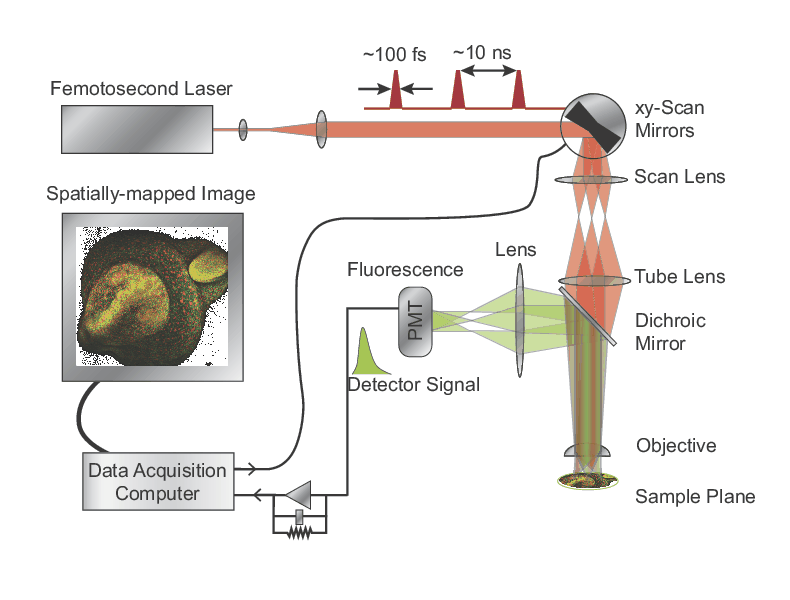
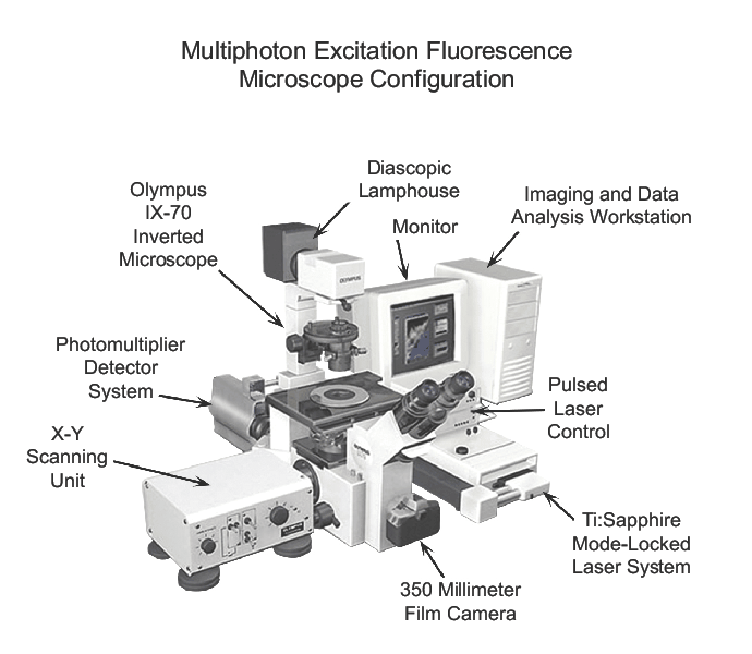
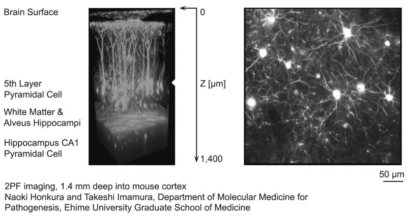
A schematic of a microscope system for 2PF is shown in Figure 3. A laser is focused to a tight spot in the specimen plane and scanned in a raster over the sample. When the laser focus overlaps with fluorescent molecules in the sample, fluorescence is generated selectively in the tiny focal volume and detected by photodetectors. The signal is spatially-mapped to generate individual pixels of an image by a data acquisition computer. The principal differences between confocal and 2PF microscopes are the laser and the fluorescence detection path. In 2PF microscopy, all fluorescent photons collected by the objective constitute useful signal as the detector pinhole is not required.

Schematic of a 2PF microscope
Figure 3. Schematic of a 2PF microscope.
Starting in the mid-1990's, the laser system of choice for 2PF was the mode-locked Ti:Sapphire laser. These lasers typically produce 100 fs pulses at an 80 MHz repetition rate with 1 W of average power and a wavelength tunable from 680 to 1050 nm. This tunability means they are ideally suited for generating 2PF for a large number of fluorescent dyes. As 2PF microscopy grew in popularity over the next 15 years, several improvements were implemented. Multiple manufacturers offered microscopes optimized for 2PF, which included improved optics for longer wavelengths and sophisticated scanners, detectors, and software. The laser sources became more robust, compact, and easy to use. For instance, the pump laser was integrated together with the Ti:Sapphire laser in a single-box system and wavelength tuning was performed through an automated computer interface. A typical layout of a commercial 2PF microscope system is shown in Figure 4.
A commercial 2PF microscope system
Figure 4. A commercial 2PF microscope system. Image courtesy of Olympus Corporation.
One challenge in the development of 2PF microscopy was to deliver 100 fs pulses to the focal plane of the sample. The pulses produced by the laser pass through multiple optics, including sophisticated multi-element microscope objectives and acousto-optic modulators where the dispersion of the materials broadens the pulses. This effect produces 200-300 fs pulses at the focus and, since two-photon excitation scales inversely with the pulse duration, this greatly reduces the intensity of the fluorescence. Fortunately, the dispersion of the materials used in the beam path can be pre-compensated with a sequence of four prisms. Automated dispersion compensation was later added to single-box laser systems.
A 3D reconstructed image of a mouse cortex up to a depth of 1.4 mm using 2PF microscopy
Figure 5. A 3D reconstructed image of a mouse cortex up to a depth of 1.4 mm using 2PF microscopy.
The penetration depth in 2PF microscopy is improved relative to one-photon excitation but is still limited to less than 1 mm in many biological samples due to scattering. Scattering decreases greatly at longer wavelengths and so there was a desire to move to longer wavelengths to improve the penetration depth. A new generation of sources that could tune from 680 nm to 1300 nm was introduced in 2011. These sources were based on NIR pump lasers and tunable oscillators. They produced similar pulse durations and average powers as the Ti:Sapphire lasers and included automated tuning and dispersion compensation in a single box. Most importantly, they nearly doubled the tuning range of Ti:Sapphire lasers, including the longer wavelengths desired for deeper penetration. As shown in Figure 5, these longer wavelengths allow imaging of a mouse hippocampus at a depth of 1.4 mm. In Figure 6, fs pulses at 980 nm were used to excite the dye enhanced green fluorescent protein (eGFP) used to label the cell membrane while pulses at 1041 nm excited the dye mCherry which labeled the cell nuclei. This demonstrates both the use of multiple wavelengths to excite different dyes and the monitoring of live samples over a period of eight hours without damage to the sample.
Embryo development monitored for eight hours in a live zebra fish embryo using 2PF microscopy
Figure 6. Embryo development monitored for eight hours in a live zebra fish embryo using 2PF microscopy.
In SHG microscopy, the fluorescent dye is not required as the second harmonic signal is generated from the sample itself. When a material is identical to its mirror image, it is said to possess inversion symmetry. SHG arises only from materials that lack inversion symmetry. For example, thin membranes can be probed with SHG microscopy. Several key endogenous protein structures, notably collagen, also give rise to intense SHG. SHG microscopy on a laser-scanning system has proven to be a powerful and unique tool for high-resolution, high-contrast, 3D studies of live cell and tissue architecture. Unlike 2PF, SHG suffers no inherent photobleaching or toxicity and does not require exogenous labels. SHG microscopy is just one of several nonlinear microscopies that do not require labeling together with others such as THG and SFG microscopy.

For nearly 20 years, the MKS Spectra-Physics MaiTai® laser has been the workhorse of bio-imaging labs around the world. It is based on traditional Ti:Sapphire technology and offers a 690 nm - 1040 nm tuning range with up to 2 W of output power and a 100 fs pulse duration. Following its initial release in 1999, it was the first laser to provide automatic wavelength tuning. Then, in 2007, it was the first laser to integrate automatic dispersion control through its DeepSee™ laser technology. In addition to its application in multiphoton microscopy, the MaiTai laser is also used in the areas of time-resolved photoluminescence, nonlinear spectroscopy, optical computed tomography, surface SHG, terahertz imaging, semiconductor metrology, materials processing, and laser amplifier seeding.

The InSight® X3™ is the third generation of MKS Spectra-Physics' industry-leading laser platform that is specifically designed for advanced multiphoton microscopy applications. The InSight X3 laser features a broad 680 nm to 1300 nm continuous, gap-free tuning range from a single source, nearly doubling the tuning range of legacy Ti:Sapphire ultrafast lasers. The system delivers high average and peak power levels across the tuning range, including the critical NIR wavelengths above 900 nm, which allow for the deepest penetration in vivo imaging. With MKS Spectra-Physics' integrated patented DeepSee™ laser technology, the industry standard dispersion pre-compensator, fs pulses are optimally delivered through a microscope to the sample for maximum fluorescence and penetration depth.


MKS Semiconductor Handbook Cover

For additional insights into photonics topics like this, download our free MKS Instruments Handbook: Principles & Applications in Photonics Technologies

Request a Handbook