Ophthalmic Surgery
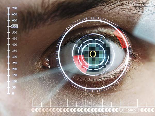
Ophthalmology is the branch of medicine that deals with the anatomy, physiology and diseases of the eye. The laser is particularly suited to ophthalmic surgery since it can provide a non-contact method of interacting with the cornea or even the interior of the eye, including the lens and the retina. Laser photocoagulation surgery is used to treat several eye diseases and has become widely used in recent decades. During this procedure, a laser is used to finely cauterize ocular blood vessels to attempt to bring about various therapeutic benefits. Treatments for diabetic retinopathy and macular degeneration have also been accomplished with several kinds of lasers including ion lasers, dye lasers, and laser diodes. Recently, two forms of ophthalmic surgery have attracted great attention: laser-assisted in situ keratomileusis (LASIK), which is used to modify the cornea and correct vision, and cataract surgery, which can facilitate replacement of the lens when it becomes cloudy. Both procedures are accomplished with the help of ultrafast pulsed laser systems, as described below.
LASIK
LASIK is the most commonly-performed interventional procedure to correct myopia (short-sightedness), hyperopia (far-sightedness), and astigmatism. It relies on optimizing the refractive properties of the cornea by removing corneal tissue through photoablation using UV ns pulses from an excimer laser. LASIK involves the creation of a thin corneal flap to give the excimer laser pulses access to deeper corneal layers. The flap is closed back to its original position after photoablation, as the preservation of the surface epithelial layer accelerates recovery. Traditionally the flap has been created mechanically using specialized mechanical cutting tools called microkeratomes. However, in recent years, these microkeratomes have been widely replaced by clinically-approved "blade-less" devices, which utilize NIR fs laser pulses for the creation of the flap.
The cornea is transparent at NIR wavelengths. However, in the focus of the beam, the peak power density can be high enough to cause laser-induced optical breakdown and photodisruption of the tissue. By using fs lasers together with a high NA objective, the damage can be spatially confined to the focal volume. The highly localized deposition of energy generates a microplasma, which can be followed by deleterious side effects such as the emission of a shock wave and the generation of cavitation bubbles . However, in contrast to ns or even ps pulses, fs laser photodisruption requires much lower pulse energies, which minimizes the range of the photodisruptive shock wave and the size of the cavitation bubbles. As a result, the collateral thermal damage zone is extremely limited since the thermal diffusion is in the sub-micron range.
The corneal flap for LASIK is created by scanning the focus of a fs laser beam at the desired depth to form a dissection plane while leaving a hinge for the flap to remain connected with the cornea. Compared to a microkeratome, the fs laser allows for flexibility with respect to the thickness of the flap. Furthermore, it enables much greater precision, which results in faster recovery times, improved visual results, and greater safety. The procedure was introduced first by Intralase, which has since been acquired by Abbott Medical Optics. Today, there are several suppliers for fs laser-based instruments, that have been clinically approved for cutting corneal flaps. The lasers deployed in these devices are DPSS or fiber lasers delivering pulse widths of a few hundred fs at repetition rates in the high kHz to low MHz range. Pulse energies can reach a few micro joules, which typically requires pulse amplification. This method for amplifying fs pulses called chirped pulse amplification was recognized with the 2018 Nobel Prize in Physics.
Flap cutting for LASIK was the first fs laser-based, clinically-approved vision correction procedure, but others have followed. Currently, there is significant focus on bringing devices to market that utilize fs pulses for high-precision interventional processes involved in the transplant of intraocular lenses (IOLs) during cataract surgery, as will be described below.
Cataract Surgery
A cataract is a clouding of the lens inside the eye causing vision loss that cannot be corrected with glasses, contact lenses, or corneal refractive surgery like LASIK. Most cataracts are associated with the aging process. Metabolic changes of the crystalline lens fibers over time lead to the development of an opacification in the lens and loss of transparency, causing impairment of vision. According to the National Eye Institute (NEI), 68.3% of Americans 80 and older had cataracts in 2010. The prevalence of cataracts in the United States is expected to grow significantly in the years ahead due, in part, to the aging of the population. In 2010, roughly 24.4 million Americans had cataracts and that number is projected to grow to 50.2 million by the year 2050, according to NEI.
Modern cataract surgery is one of the safest and most effective surgical procedures performed today. More than 3 million cataract surgeries are performed in the United States every year, with the majority of these procedures producing excellent visual outcomes. In cataract surgery, the lens inside the eye that has become cloudy is removed and replaced with an artificial lens (or IOL) to restore clear vision. Today, the procedure is typically performed on an outpatient basis.
There are two main types of surgical procedures in use today. The first procedure is phacoemulsification (or phaco) and the second is extracapsular cataract extraction (ECCE). Phaco is the most commonly-performed cataract procedure in the developed world while ECCE, which is less expensive, is more frequently performed in developing countries. Phaco involves the use of a high-frequency (40 kHz) ultrasound device that breaks up the cloudy lens into small pieces, which are then gently removed from the eye with suction. This can be performed with smaller incisions than previous surgical techniques for cataract removal, promoting faster healing and reducing the risk of cataract surgery complications. After all remnants of the cloudy lens have been removed from the eye, the cataract surgeon inserts a clear IOL, positioning it securely behind the iris and pupil, in the same location the natural lens occupied. The surgeon then completes the cataract removal and IOL implantation procedure by closing the incision in the eye.
Recently, a number of fs lasers, similar to the lasers used to create the corneal flap in LASIK, has been approved by the FDA for use in cataract surgery performed in the United States. The main advantages are standardized corneal incisions, perfectly centered, round openings of the lens capsule (capsulorhexis), and lens nucleus fragmentation even in eyes with hard cataracts. Laser cataract surgery is relatively new and significantly increases cataract surgery cost, in part, due to the cost of the laser. In the United States, private insurance and Medicare cover most if not all of the associated costs. There are currently at least five companies with fs laser-based cataract surgery platforms, including Alcon LenSX, LENSAR, Abbott Medical Optics (OptiMedica), Bausch & Lomb Technolas, and Ziemer. Fs lasers have gained approval for multiple steps in cataract surgery, which reduces the need for surgical blades and other hand-held tools. These surgical steps include creating corneal incisions to allow the surgeon access to the lens, performing so-called limbal relaxing incisions to reduce or eliminate pre-existing astigmatism, removing the anterior capsule of the lens, and fragmenting the cataract so lower phaco energy is required to break it up.
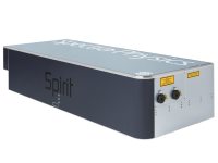
The MKS Spectra-Physics Spirit fs laser is a one-box amplifier which delivers 400 fs pulses with 4 W of average power at 1040 nm. With up to 40 µJ of energy, this laser has been extensively deployed for LASIK flap cutting and has the capability of being implemented for approved cataract surgical steps.
Future Directions of Ophthalmic Surgery
In LASIK, NIR fs lasers are used to cut the flap in the cornea while an excimer laser is used to ablate the exposed portion. It would be desirable if both processes could be achieved with a single laser. This has recently been achieved with the introduction of the Refractive Lenticule Extraction Small Incision Lenticule Extraction (ReLEx SMILE) procedure developed by Carl Zeiss Meditec. The procedure uses a single fs laser system to cut a small lens-shaped piece (known as lenticule) within the corneal stroma and to create a small incision along the periphery of this lenticule to remove it through the incision. In a similar vein to LASIK, cataract surgery requires both an ultrafast laser to make the incision in the cornea and an ultrasound source to fragment the lens material that will be removed. Initial results have been announced that use a fs laser for both the incision and the photofragmentation of the lens.
Presbyopia is another age-related condition of the eye. It is the normal loss of near-focusing ability that occurs with age with the majority noticing the effects of presbyopia sometime after the age of 40. This typically manifests in difficulty in reading small print such as text messages on a smartphone. According to a global report on presbyopia issued in 2012 by Market Scope, worldwide, an estimated 1.3 billion people had presbyopia in 2011. This number is expected to increase to 2.1 billion by 2020. Currently, the most common treatments are the use of reading glasses or contact lenses. A special type of contact lens correction for presbyopia is monovision, in which one eye wears a distance prescription, and the other wears a prescription for near vision. The brain learns to favor one eye or the other for different tasks. There also are surgical treatments such as the implantation of a corneal inlay. A corneal inlay is typically implanted in the cornea of the person's non-dominant eye. Like monovision, this approach helps increase the depth of focus of the treated eye and reduces the need for reading glasses without significantly affecting the quality of distance vision. The sole corneal inlay that is approved by the FDA is the KAMRA inlay. Since virtually every person will get presbyopia given a long enough life span, several medical companies are working on developing a laser-based treatment for this ever-expanding market.
There is an intriguing application for ultrafast lasers currently under investigation with the potential to impact both cataract surgery and vision correction. In both LASIK and cataract surgery, ultrafast lasers cut material. This process of micro-machining the cornea requires amplified fs laser systems with pulse energies of several micro joules. Lower energy fs lasers, instead of cutting, only modify the index of refraction in transparent materials. Through the process of multiphoton absorption, the refractive index modification is localized in three dimensions due to the nonlinear nature of the process. Initial work has focused on using this approach to modify the refractive index in either contact lenses or the IOLs that are implanted during cataract surgery. Researchers have been able to modify the index and write Fresnel lens structures with an optical power of several diopters into the materials used for contacts. Clerio Vision is pursuing this process with unamplified laser oscillators that produce fs pulses with nJ energies at 800 nm, while Perfect Lens is using amplified fs pulses with .J energies at 515 nm.
More exciting, and more challenging, is to write the index change directly into the cornea of the patient. It has been reported that only 2% of those needing refractive correction of some sort have had LASIK. Thus, a less invasive procedure might find wide adoption. Fs pulses at a wavelength of 405 nm with nanojoules of energy are required for this approach. The wavelength is chosen to optimize the index change in the cornea while protecting the retina. Currently, these pulses are produced by generating the second harmonic of fs oscillators operating at 800 nm. Animal studies have been underway for several years with encouraging results. Successful implementation will depend on many factors, including longevity of the correction, successful trials and FDA approval, as well as the development of lower-cost, dedicated fs laser systems.
Ophthalmic Surgery Products
For additional insights into photonics topics like this, download our free MKS Instruments Handbook: Principles & Applications in Photonics Technologies
Request a Handbook