Currently, there are not many fluorescent dyes where the two-photon excitation wavelength overlaps with these optimal wavelengths. However, there are dyes with a spectral overlap of their three-photon absorption in these low loss windows. Some of these dyes are the same as those used for 2PF microscopy at 900 nm, e.g., green fluorescent protein (GFP) based probes, and 1100 nm, e.g. red fluorescent protein (RFP) based probes. Over the last decade, commercial manufacturers have improved the transmission of their microscopes for longer wavelength excitation.
One remaining challenge is that the cross-section (or the probability of excitation) for 3PF is several orders of magnitude smaller than for 2PF, requiring lasers with much higher peak powers for efficient excitation. However, to prevent damage at the surface of samples, the average power must be limited. Thus, to achieve the required peak powers, laser systems that produce 100 fs pulses with higher energy but with lower repetition rates (to maintain modest average power) are required. A typical laser for 3PF microscopy produces 100 fs pulses every 1 µs (a repetition rate of 1 MHz). Therefore, 1 W of average power gives a peak power of 10 MW with the laser on only 0.00001% of the time. Laser systems that produce ultrashort pulses at this high energy are based on ultrafast amplifier systems that operate at a wavelength of 1 µm. To shift the wavelength to the target wavelengths of 1.3 µm and 1.7 µm, additional nonlinear conversion schemes are required including SHG and OPG.
In THG microscopy, the fluorescent dye is omitted and the third harmonic signal is generated from the sample itself. In a homogenous sample, the THG signal from above the focus cancels the signal generated below the focus due to phase matching. Thus, a third harmonic signal is only generated when the focus is close to a gradient in the refractive index. As a result, THG microscopy is particularly well-suited for 3D label-free imaging of transparent specimens where the membrane or other interface is of interest.
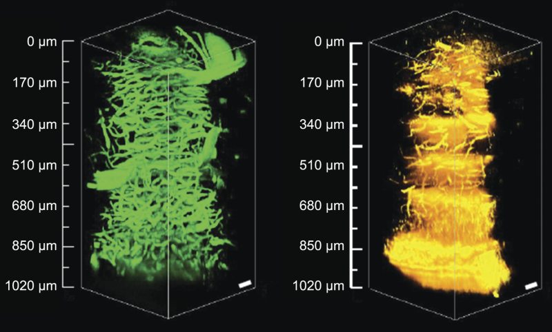
When a combination of techniques is utilized to interrogate a single sample, it is referred to as multi-modal imaging. Using multiple techniques allows investigators to look at different features in the same sample as illustrated in Figure 3, which shows a 3D reconstruction of a mouse brain cerebellum. Fs pulses at 1.3 µm are used to excite the tissue, which generates 3PF in fluorescein-labeled blood vessels while the same wavelength also produces a THG signal in different portions of the cerebellum, i.e., myelinate fibers.
 Over 8,000 products in-stock! & FREE 2-Day shipping on all web orders!* Learn More FREE T-Shirt with orders $250+ Details
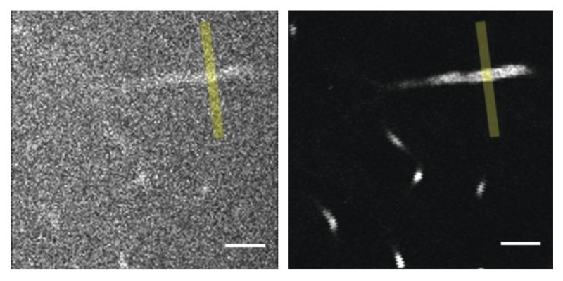

 Ultra-High Velocity